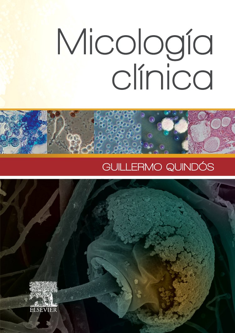
Quindós Andrés G. Micología clínica. Elsevier España, Barcelona. 2015. Abstract: La Micología clínica es una especialidad en constante evolución que estudia las infecciones causadas por los hongos, llamadas micosis. La frecuencia y la importancia de las micosis es cada vez mayor en las personas con estados de inmunodeficiencia o que sufren enfermedades debilitantes y graves. Dentro de la Micología clínica nos enfrentamos a nuevos e importantes retos, porque las especies fúngicas tanto clásicas como nuevas que causan estas enfermedades son cada vez más numerosas y diversas. El diagnóstico de las micosis invasoras es complicado, pues en muchas ocasiones su presentación clínica es indistinguible de la de aquellas enfermedades producidas por otros patógenos, bacterianos, víricos o protozoarios. El tratamiento, tanto empírico como dirigido, es difícil de establecer, y resulta necesaria una profunda y extensa investigación clínica que permita reducir la inaceptablemente alta mortalidad de muchas de estas micosis invasoras.

Quindós G, Marcos Arias C, Eraso E, Guarro J. Trichosporon, Magnusiomyces and Geotrichum. En: Paterson RRM (Eds.). Molecular Biology of Food and Water Borne Mycotoxigenic and Mycotic Fungi. CRC Press, Taylor & Francis Group. Boca Ratón, Florida, EE UU. 2015. Abstract: Trichosporon, Magnusiomyces, and Geotrichum are ubiquitous fungi in nature, closely associated with plants and animals, and can be part of the human microbiota. Moreover, these fungi cause human disease when they cause deep infections with a high mortality (40%–80%), although most isolates show a low virulence and infections are superficial. The terms trichosporonosis and geotrichosis refer to those mycoses caused by Trichosporon, and Geotrichum and Magnusiomyces, respectively. Invasive mycoses caused by these fungi are infrequent in the general population; however, the increase in the number of immunodeficient patients and use of antifungal prophylaxis has converted them into emergent opportunistic agents causing severe nosocomial infections worldwide. Moreover, these infections have also been observed in patients suffering from cancer and in critically ill patients exposed to multiple invasive medical procedures. The ability of Trichosporon, Magnusiomyces, and Geotrichum to adhere to and develop biofilms on implanted devices may account for persistence and dissemination, since this ability promotes escape from host immune responses and resistance to antifungal drugs. In addition, the presence of glucuronoxylomannan in the cell wall of Trichosporon and the ability of these fungi to produce hydrolytic enzymes are additional virulence factors related to the progression of infections.
Quindós Andrés G. Risks groups in acquiring fungal infections. Chapter 6. Fungal specificities in Environmental Mycology. En: Viegas C, Pinhero AC, Sabino R, Viegas S, Brandão J, Veríssimo C (Eds.). Environmental Mycology in Public Health. Indoor Fungi and Mycotoxins Risk Assessment and Management. Academic Press / Elsevier, Whaltam, Maryland, EE UU. 2015. Abstract: The epidemiology of mycoses is in a continuous flux associated with medical and surgical advances. Most people in regular contact with fungi do not suffer any mycoses, despite the fungal ubiquity. However, there is a growing population with underlying diseases or predisposing factors that favor invasive mycoses. Many fungal infections are acquired by inhalation of, contact with, or ingestion of fungal propagules or by fungi entering the bloodstream through needles or catheters, etc. Invasive candidiasis is the most frequent mycosis but invasive aspergillosis can be predominant in bone marrow transplant recipients. Other fungi, such as Pneumocystis, Cryptococcus, Fusarium, and Rhizopus, can cause devastating illnesses. Moreover, there are significant epidemiological variations between countries and between hospitals in the same country that must be related both to local characteristics of the disease and its risk factors and to differences in the practices of the various medical and surgical services. Patients at higher risk are those admitted to intensive care units; using prostheses, catheters, or other intravenous devices; or receiving various immunosuppressant treatments or antineoplastic chemotherapy and transplant recipients. In addition, opportunistic mycoses can be associated with human immunodeficiency virus infection. Mortality attributed to fungal diseases is high, ranging from 30% in invasive candidiasis to 90–100% in some clinical presentations of mucormycosis. The great complexity of patients presenting important risks and the growing diversity of pathogenic fungi are major diagnostic and therapeutic challenges.
Quindós G, Salavert M. Zigomicosis (Mucormicosis). Capítulo 184. En: Vesga O, Vélez LA, Leiderman E, Restrepo A (Eds.). Fundamentos de Medicina: Enfermedades Infecciosas del Homo sapiens. Corporación para Investigaciones Biológicas (CIB), Medellín, Colombia. 2015. Abstract: Las mucormicosis son las enfermedades infecciosas causadas por los hongos filamentosos denominados Mucorales. El término zigomicosis o cigomicosis abarca un rango más amplio de micosis porque incluye tanto las mucormicosis como las enfermedades causadas por los hongos Entomophthorales, denominadas entomoftoromicosis. Sin embargo, los sinónimos antiguos de zigomicosis, como ficomicosis e hifomicosis, se deben considerar obsoletos. Como ficomicosis se denominaban a las infecciones causadas por los zigomicetos y por otros hongos filamentosos, como Pythium, muchos de los cuales están ahora clasificados en otros reinos diferentes al reino Fungi, como el reino Chromista. En este capítulo se emplearán los términos mucormicosis y zigomicosis como sinónimos para evitar las controversias relacionadas con la mayor familiaridad del término mucormicosis para los médicos clínicos y la preferencia del término zigomicosis para la mayoría de los micólogos médicos. Finalmente, se utilizará el término entomoftoramicosis de forma restringida cuando se trate de las micosis causadas por los géneros Basidiobolus o Conidiobolus.
Quindós G, Eraso E, Ezpeleta G, Pemán J, Sanchez Reus F. State of the art in the laboratory methods for the diagnosis of invasive fungal diseases. En: Kon K & Rai M (Eds.). Microbiology for Surgical Infections: Diagnosis, Prognosis and Treatment. Academic Press / Elsevier. Whaltam, Maryland, EE UU. 2014. Abstract: Invasive fungal diseases (IFDs) possess a great clinical relevance because of their growing incidence, morbidity and mortality in oncohematological and surgical patients. Immunosuppression and breakdown of anatomical barriers are major risk factors and most at-risk patients are in surgery services, AIDS clinics, intensive care units (ICU), or transplantation and oncology units. Invasive candidiasis (IC) and to a lesser extent pulmonary and disseminated aspergillosis, pulmonary pneumocystosis and meningeal cryptococcosis are the most frequent IFDs. Organ transplant recipients form an important group of at-risk patients, with the type of transplanted organ causing major differences in etiology; thus, aspergillosis and other filamentous fungi infections are common in bone marrow transplants. However, IFDs are dominated by Candida in the first month after solid organ transplantation, in a manner usually related to surgical issues and nosocomial risk factors. Thus, anastomotic leaks, early graft failure, reoperation, and central venous catheter-associated candidemia are common. In the absence of complications of transplant, IFD by filamentous fungi are uncommon. After the first month, intense immunosuppression methods, such as direct microscopy, histology or culture of clinical specimens, are not sensitive or specific enough, and the results are often available too late to be clinically useful. This delay habitually enables the IFD to progress to the point where antifungal therapy becomes ineffective, thus there is a close relationship between early diagnosis, prompt treatment, and lower mortality. The prompt initiation of antifungal therapy is critical for improving the IFD outcome, and in many cases this has led to empirical treatment. Early mycological diagnosis is the cornerstone of a prompt and appropriate treatment, and for improving the survival of patients. Diagnostic methods are in continual evolution, and more rapid and sensitive methods have been developed, such as the detection of biomarkers or surrogate markers, e.g., 1,3-β-D-glucan (BG), glucuronoxylomannan (GXM), galactomannan (GM), mannan (MN), antimannan and anti-Candida germtube antibodies (CAGTA), and fungal DNA detection by polymerase chain reaction (PCR). However, many of these approaches are unfamiliar and they are generally not available in most clinical settings.
Views: 0
